Печать
ANTI-ARFGAP2
Кат. №: HPA018152-100UL
Производитель: Sigma-Aldrich
Кол-во:
Цена по запросу
Товар оформляется под заказ
Печать
ANTI-ARFGAP2
Кат. №: HPA018152-100UL
Производитель: Sigma-Aldrich
Кол-во:
Цена по запросу
Товар оформляется под заказ
Кол-во:
Цена по запросу
Товар оформляется под заказ
Description_x000D_
General description_x000D_
The gene ADP-ribosylation factor GTPase-activating protein-2 (ARFGAP2) has been mapped to human chromosome 11p11.2. ARFGAPs form a family of proteins sharing a conserved catalytic domain which include a zinc finger motif, and they differ in the non-catalytic domains. Human genome contains sixteen genes coding for ARFGAPs. Fluorescent protein tagging showed ARFGAP2 localization on the Golgi complex and punctuate structures in the cytoplasm identified as endoplasmic reticulum-Golgi intermediate compartments/vesicular-tubular compartments._x000D_
Immunogen_x000D_
ADP-ribosylation factor GTPase-activating protein 2 recombinant protein epitope signature tag (PrEST)_x000D_
Application_x000D_
All Prestige Antibodies Powered by Atlas Antibodies are developed and validated by the Human Protein Atlas (HPA) project (www.proteinatlas.org)and as a result, are supported by the most extensive characterization in the industry._x000D_
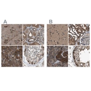
The Human Protein Atlas project can be subdivided into three efforts: Human Tissue Atlas, Cancer Atlas, and Human Cell Atlas. The antibodies that have been generated in support of the Tissue and Cancer Atlas projects have been tested by immunohistochemistry against hundreds of normal and disease tissues and through the recent efforts of the Human Cell Atlas project, many have been characterized by immunofluorescence to map the human proteome not only at the tissue level but now at the subcellular level. These images and the collection of this vast data set can be viewed on the Human Protein Atlas (HPA) site by clicking on the Image Gallery link. To view these protocols and other useful information about Prestige Antibodies and the HPA, visit sigma.com/prestige._x000D_
Physical form_x000D_
Solution in phosphate-buffered saline, pH 7.2, containing 40% glycerol and 0.02% sodium azide_x000D_
Disclaimer_x000D_
Unless otherwise stated in our catalog or other company documentation accompanying the product(s), our products are intended for research use only and are not to be used for any other purpose, which includes but is not limited to, unauthorized commercial uses, in vitro diagnostic uses, ex vivo or in vivo therapeutic uses or any type of consumption or application to humans or animals._x000D_
Legal Information_x000D_
Prestige Antibodies is a registered trademark of Sigma-Aldrich Co. LLC_x000D_
Biochem/physiol Actions_x000D_
ADP-ribosylation factor GTPase-activating protein-2 (ARFGAP2) is shown to associate with COP-I-coated vesicles produced in-vitro. Additionally, it is shown to be involved in the COP-I-dependent Golgi-to-endoplasmic reticulum trafficking of a model cargo, cholera toxin. ARFGAP1, 2 and 3 together are essential for COP-I-mediated trafficking in mammalian cells. Triple knockout of ARFGAP1/2/3 in HeLa cells result in cell death. Single knockdown of ARFGAP2 in HeLa cells using siRNA approach results in Golgi unstacking and cisternal shortening. Brefeldin A treatment results in re-distribution of the ARFGAP2 from the Golgi complex to the cytosol._x000D_
Features and Benefits_x000D_
Prestige Antibodies® are highly characterized and extensively validated antibodies with the added benefit of all available characterization data for each target being accessible via the Human Protein Atlas portal linked just below the product name at the top of this page. The uniqueness and low cross-reactivity of the Prestige Antibodies® to other proteins are due to a thorough selection of antigen regions, affinity purification, and stringent selection. Prestige antigen controls are available for every corresponding Prestige Antibody and can be found in the linkage section._x000D_
Every Prestige Antibody is tested in the following ways:_x000D_
• IHC tissue array of 44 normal human tissues and 20 of the most common cancer type tissues._x000D_
• Protein array of 364 human recombinant protein fragments._x000D_
• Validated for multiple commonly used applications such as IHC (Immunohistochemistry), IF (Immunofluorescence), and WB (Western Blot)_x000D_
Linkage_x000D_
Corresponding Antigen APREST73727.
Related Categories
AQ-AZ, AR-AR, Alphabetical Index, Antibodies, Prestige Antibodies, Prestige Polyclonal Antibodies, Primary AntibodiesMore... product line
Дорогой клиент, на сайте внедрена нейросеть для сбора информации о товаре. Это может привести к незначительным расхождениям в характеристиках продукции.