Печать
Anti-p38 MAP Kinase (341-360 1PC X 200UL
Кат. №: 506123-200UL
Производитель: Sigma-Aldrich
Кол-во:
Цена по запросу
Товар оформляется под заказ
Печать
Anti-p38 MAP Kinase (341-360 1PC X 200UL
Кат. №: 506123-200UL
Производитель: Sigma-Aldrich
Кол-во:
Цена по запросу
Товар оформляется под заказ
Кол-во:
Цена по запросу
Товар оформляется под заказ
Description
General description
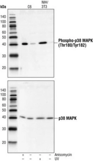
Protein A and immunoaffinity purified rabbit polyclonal antibody. Recognizes the ~38 kDa p38 MAPK protein.
Recognizes the ~38 kDa p38 MAPK protein.
Antibody Target Gene Symbol: MAPK14
• Target Synonym: CRK1, CSBP, CSBP1, CSBP2, CSPB1, EXIP, Hog, MAPK p38, MGC102436, MGC105413, MXI2, P38, P38 KINASE, P38 Map Kinase, p38 Mapk alpha, P38-ALPHA, p38-RK, p38/Hog1, p38/Mpk2, P38/RK, p38a, p38Hog, p38MAPK, PRKM14, PRKM15, RK, SAPK2A
• Entrez Gene Name: mitogen-activated protein kinase 14
• Hu Entrez ID: 1432
• Mu Entrez ID: 26416
• Rat Entrez ID: 81649
This Anti-p38 MAP Kinase (341-360) Rabbit pAb is validated for use in Flow Cytometry, Immunoblotting, Paraffin Sections for the detection of p38 MAP Kinase (341-360).
Immunogen
Human
a synthetic peptide (TYDEYISFVPPPLDQEEMES) corresponding to amino acids 341-360 of human p38 MAP kinase
Application
Flow Cytometry (1:25)
Immunoblotting (1:1000)
Paraffin Sections (1:50, heat pretreatment required, see comments)
Physical form
In 150 mM NaCl, 10 mM HEPES, 50% glycerol, 0.01% BSA, pH 7.5.
Reconstitution
Following initial thaw, aliquot and freeze (-20°C).
Legal Information
TWEEN is a registered trademark of Croda International PLC
Packaging
200 μL in Plastic ampoule
Warning
Toxicity: Standard Handling (A)
Other Notes
Pretreat paraffin sections by heating tissue in 10 mM citrate buffer, pH 6.0 for 1 min at high power followed by 9 min at medium power; keep the slides fully immersed and maintain the temperature at or just below boiling; cool the slides for 20 min at room temperature prior to staining. Variables associated with assay conditions will dictate the proper working dilution.
Recommended Protocol for Immunoblotting
Solutions and Reagents
• Transfer Buffer: 25 mM Tris base, 0.2 M glycine, 20% methanol, pH 8.5.
• SDS Sample Buffer: 62.5 mM Tris-HCl, pH 6.8, 2% SDS, 10% glycerol, 50 mM DTT, 0.1% bromophenol blue.
• 10X TBS (Tris-buffered saline): To prepare 1 liter, 24.2 g Tris base, 80 g NaCl, adjust pH to 7.6 with HCl. Dilute 1:10 for use.
• Blocking Buffer: 1X TBS, 0.1% Tween®-20 detergent with 5% non-fat dry milk.
• Primary Antibody Dilution Buffer: 1X TBS, 0.1% Tween-20 detergent with 5% BSA
• Wash Buffer (TBST): 1X TBS, 0.1% Tween-20 detergent
Blotting Membrane
Nitrocellulose or PVDF membranes may be used.
Protein Blotting
1. Lyse cells by adding 100 ml SDS Sample Buffer and immediately scrape the cells off the plate and transfer the extract to a microfuge tube. Keep on ice.
2. Sonicate for 2 s to shear DNA and reduce sample viscosity.
3. Heat sample to 95-100°C for 5 min. Cool on ice.
4. Microcentrifuge for 5 min.
5. Load 20 ml onto SDS-PAGE gel (10 cm x 10 cm).
6. Electrotransfer to nitrocellulose membrane.
As controls, we recommend using 15 ml of phosphorylated and nonphosphorylated C-6 glioma cell extracts.
Membrane Blocking, Gel and Antibody Incubations
1. After transfer, wash membrane with 25 ml TBS for 5 min at room temperature.
2. Incubate membrane in 25 ml of Blocking Buffer for 1-3 h at room temperature or overnight at 4°C.
3. Wash 3 times for 5 min each with 15 ml TBST.
4. Incubate membrane and primary antibody (at the appropriate dilution) in 10 ml Primary Antibody Dilution Buffer with gentle agitation overnight at 4°C.
5. Wash 3 times for 5 min each with 15 ml TBST.
6. Incubate membrane with conjugated secondary antibody at the appropriate dilution in 10 ml Blocking Buffer with gentle agitation for 1 h at room temperature.
7. Wash membrane as in step 5.
Detection of Proteins
Chemiluminescence.
Raingeaud, J., et al. 1995. J. Biol. Chem.270, 7420.
Zervos, A.S., et al. 1995. Proc. Natl. Acad. Sci. USA92, 10531.
Han, J., et al. 1994. Science265, 808.
Lee, J.C., et al. 1994. Nature372, 739.
Rouse, J., et al. 1994. Cell78, 1027.
Related Categories
Alphabetical Index, Antibodies, P-PA, Primary Antibodies clone
Дорогой клиент, на сайте внедрена нейросеть для сбора информации о товаре. Это может привести к незначительным расхождениям в характеристиках продукции.