Печать
Anti-Pan-Neuronal Neurofilam 1PC X 100UL
Кат. №: NE1017-100UL
Производитель: Sigma-Aldrich
Кол-во:
Цена по запросу
Товар оформляется под заказ
Печать
Anti-Pan-Neuronal Neurofilam 1PC X 100UL
Кат. №: NE1017-100UL
Производитель: Sigma-Aldrich
Кол-во:
Цена по запросу
Товар оформляется под заказ
Кол-во:
Цена по запросу
Товар оформляется под заказ
Description
General description
Mouse monoclonal antibody supplied as undiluted ascites. Recognizes the ~180-200 kDa pan-neuronal neurofilament marker protein.
Recognizes ~180-200 kDa pan-neuronal neurofilament marker in rat central nervous system cytoskeletal preparations.
This Anti-Pan-Neuronal Neurofilament Marker Mouse mAb (SMI-311) is validated for use in ELISA, Frozen Sections, WB, ICC, Paraffin Sections for the detection of Pan-Neuronal Neurofilament Marker.
Immunogen
Rat
homogenized hypothalami extracted from Fischer 344 rat brain
Application
ELISA (1:1000)
Frozen Sections (1:1000, see comments)
Immunoblotting (1:1000)
Immunocytochemistry (1:1000, see comments)
Paraffin Sections (1:1000, heat pre-treatment required, see comments)
Physical form
Undiluted ascites.
Reconstitution
Following initial thaw, aliquot and freeze (-20°C).
Packaging
100 μL in Glass bottle
Warning
Toxicity: Standard Handling (A)
Analysis Note
Positive Control
Rat brain or rat CNS cytoskeletal preparations
Other Notes
Agostino, A., et al. 2003. Hum. Mol. Genet.12, 399.
Tsunoda, I., et al. 2003. Am. J. Pathol.162, 1259.
Ulfig, N., et al. 1998. Cell Tissue Res.291, 433.
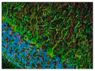
This antibody cocktail was selected to provide a specific marker for neurons in tissue sections and cultured cells. In contrast to individual anti-nonphosphoneurofilament antibodies that identify different subsets of neurons, this cocktail is a convenient marker for neurons in general and their differentiation from non-neuronal cells. Also useful as an early marker of neuronal migration and differentiation in human fetal development, yielding Golgi-like images without the disadvantages of the lack of selectivity and poor specificity of the Golgi technique. Can be used to trace the "inside-out gradient" of neuron production and differentiation in specifically delineating cell bodies and dendrites. Certain pathologic conditions, such as malnutrition, affect the SMI-311-visualized soma size and dendritic arborization. Tissues and cultured cells can be fixed in a variety of paraformaldehyde- or formaldehyde-containing fixatives, including Bouin′s fixative. This antibody does not react well with tissues or cells fixed in glutaraldehyde/paraformaldehyde. For staining paraffin sections it is recommended that de-paraffinized tissues be autoclaved in dH2O for 10 min to expose the epitope. Trypsin pre-treatment will abolish reactivity. Post-fixation in cold methanol or methanol/hydrogen peroxide facilitates access of the antibody to the neurons in frozen sections and thick tissues sections that have been fixed in 4% paraformaldehyde or cultured cells. For tissues that have not been paraffin-embedded and have been stored for long periods of time in formaldehyde fixatives, it is recommended that the tissues (up to 0.5 cm thick) be boiled in Tris-buffered saline, pH 9.0 for 15 min prior to sectioning. Antibody should be titrated for optimal results in individual systems.
Related Categories
Alphabetical Index, Antibodies, P-PA, Primary Antibodies clone
Дорогой клиент, на сайте внедрена нейросеть для сбора информации о товаре. Это может привести к незначительным расхождениям в характеристиках продукции.