Печать
Anti-Tau-1, clone PC1C6 (mouse monoc
Кат. №: MAB3420-25UG
Производитель: Sigma-Aldrich
Кол-во:
Цена по запросу
Товар оформляется под заказ
Печать
Anti-Tau-1, clone PC1C6 (mouse monoc
Кат. №: MAB3420-25UG
Производитель: Sigma-Aldrich
Кол-во:
Цена по запросу
Товар оформляется под заказ
Кол-во:
Цена по запросу
Товар оформляется под заказ
Description
General description
Tau, a microtubulebinding protein which serves to stabilize microtubules in growing axons, is found to be hyperphosphorylated in paired helical filaments (PHF), the major fibrous component of neurofibrillary lesions associated with Alzheimer’s disease. Hyperphosphorylation of Tau is thought to be the critical event leading to the assembly of PHF. Six Tau protein isoforms have been identified, all of which are phosphorylated by glycogen synthase kinase 3 (GSK 3). Cellular and subcellular localization: In situ, anti-tau-1 has a stringent specificity for the axons of neurons. The antibody does not stain the cell bodies or dendrites of neurons, nor does it stain any other cell type (4). However, this in vivo intracellular specificity is not maintained in culture: anti-tau-1 stains the axon, cell bodies, and dendrites of rat hippocampal neurons grown in culture (5). The specificity of anti-tau-1 was originally thought to represent the restricted expression of tau to axons. Later studies revealed that this specificity is dependant on the state of phosphorylation. In dephosphorylated samples (samples treated with alkaline phosphatase) anti-tau-1 stains astrocytes, perineuronal glial cells, and the axons, cell bodies and dendrites of neurons, while in untreated samples, anti-tau-1 stains only axons (6). (The epitope recognized by anti-tau-1 is probably at or near a phosphorylated site.)
Specificity
Binds to all known electrophoretic species of tau in human, rat and bovine brain (one-dimensional SDS-PAGE). However there is some unphosphorylated bias with clone PC1C6 as it seem to recognize only dephosphorylated serine sites at 195, 198, 199, and 202 {Szendrei, et al 1993; http://www.ncbi.nlm.nih.gov/entrez/query.fcgi-cmd=Retrieve&db=pubmed&dopt=Abstract&list_uids=7680727}. Also see Billingsley & Kincaid, 1997 Biochem J 323:577-591 for additional mapping information on PC1C6.
Immunogen
Purified denatured bovine microtubule associated proteins.
Application
Anti-Tau-1 Antibody, clone PC1C6 is an antibody against Tau-1 for use in IH & WB with more than 65 product citations.
Research Category
Neuroscience
Research Sub Category
Neurodegenerative Diseases
Western blot: Bovine brain microtubule proteins purified by two cycles of assembly and disassembly (9) are dissolved in SDS-PAGE sample buffer. Five micrograms of the microtuble preparation per lane is loaded onto a 4% to 20% SDS-PAGE gradient gel along side molecular weight markers (14.3 - 200 kD). After separation by electrophoresis, the proteins are blotted onto nitrocellulose. Tau is detected as a series of 5 bands (52-68 kD) with approximately 5 ng/mL of anti-tau1.
Immunofluorescence: A 1:1000 dilution of this antibody detected Tau in mouse primary neurons. (Basnet, N., et al. (2018). Nat. Cell Biol. 20(10); 1172-1180.
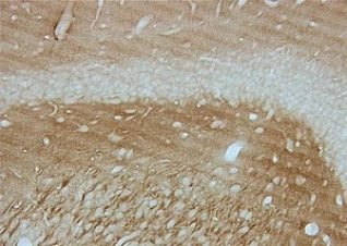
Immunohistochemistry: 5 μg/mL; stains axons in tissue primarily, however in culture Tau expression is not restricted to just axons.
Optimal working dilutions must be determined by end user.
Immunohistochemistry Protocol
Dephosphorylation of tissue sections (optional)
Dephosphorylation with alkaline phosphatase is recommended for staining neurofibrillary tangles in Alzheimer′s brain tissue with anti-tau-1 (6). This treatment changes the staining pattern of anti-tau-1 to include cell bodies, dendrites and axons of neurons. In untreated samples, anti-tau-1 stains axons only.
1. Incubate tissue sections at +32°C for 2.5 hours with constant agitation in the following solution: 100 mM Tris-HCl, pH 8.0; 130 units/mL alkaline phosphatase, 1 mM PMSF, 10 μg/mL pepstatin and 10 μg/mL leupeptin.
2. Rinse sections twice, 3 min per rinse, with 100 mM Tris-HCl, pH 8.0.
Anti-tau-1 staining
1. Block non-specific binding by incubating sections in PBS containing 1% (v/v) normal animal serum, and 0.03% (w/v) Triton X-100. The animal serum should be from the same species as the secondary antibody.
2. Rinse 3 times with PBS, 3 min per rinse.
3. Incubate sections with anti-tau-1, approximately 5 μg/mL, diluted in PBS containing 1% (v/v) normal animal serum.
4. Wash with PBS, changing the solution 3 times over a 3 min period.
5. Detect with a standard secondary antibody detection system (10-13).
Target description
5 bands (52–68 kDa)
Linkage
Replaces: AB1512
Physical form
0.02M phosphate buffer, pH 7.6, 0.25M NaCl, and 0.1% sodium azide
Format: Purified
Protein A Purfied
Storage and Stability
Maintain for 1 year at -20°C from date of shipment. Aliquot to avoid repeated freezing and thawing. For maximum recovery of product, centrifuge the original vial after thawing and prior to removing the cap.
Analysis Note
Control
Alzheimer′s brain tissue (dephosphorylation with alkaline phosphatase is recommended for staining neurofibrillary tangles in Alzheimer’s brain tissue) or human T98G glioblastoma cells
Other Notes
Concentration: Please refer to the Certificate of Analysis for the lot-specific concentration.
Disclaimer
Unless otherwise stated in our catalog or other company documentation accompanying the product(s), our products are intended for research use only and are not to be used for any other purpose, which includes but is not limited to, unauthorized commercial uses, in vitro diagnostic uses, ex vivo or in vivo therapeutic uses or any type of consumption or application to humans or animals.
Related Categories
Antibodies, Antibodies for Neurobiology, Antibodies to Neural Proteins, Miscellaneous, Neuroscience,
Primary Antibodies
More... Quality Level
Дорогой клиент, на сайте внедрена нейросеть для сбора информации о товаре. Это может привести к незначительным расхождениям в характеристиках продукции.